


Fujifilm Supria 32-Slice CT Scanner
Lets have a discussion, independent finance and extended service warranties available
The Fujifilm Supria 32 slice CT scanner offers high-resolution imaging with submillimeter slice capabilities, utilizing 0.625mm x 16 channel scanning
It features Intelli IP technology for enhanced image quality and noise reduction
The compact design includes a 75cm gantry aperture for easy patient access making it suitable for various clinical applications.
Lead time: 12-16 weeks
Description
Fujifilm Supria-32 Slice CT Scanner
Where compactness and affordability go hand in hand – Fujifilm Supria-32 Slice CT Scanner has a more cost effective setup cost therefore, your asset will pay its self off faster.
One system installed in an Auckland veterinary hospital – site visits can be arrange for interested parties.
The Fujifilm Supria 32 slice CT scanner is designed to deliver high-quality imaging with efficiency and patient comfort in mind. It features submillimeter slice imaging for high-resolution images, utilising 0.625mm x 16 channel scanning 1
- This allows for smooth 3D and multi-planar reconstruction (MPR) images
- The Supria 32 also incorporates Intelli IP, a powerful iterative reconstruction technology that enhances image quality while reducing noise
- Its compact design fits into small spaces and includes a 75cm gantry aperture for easy patient access
- Additionally, the system supports various scanning modes, including axial, helical, and dynamic scans, making it versatile for a wide range of clinical applications
“Supria” meets Healthcare Needs.

With the aging of society around the world, the demand for reducing the physical burden on patients is increasing at medical fields.
On the other hand, there is a demand for optimal and efficient hospital management, and it is also required to respond to various healthcare needs.
We will solve the challenges being faced in the medical practice with “Supria”, which can meet the healthcare needs of present and future.
“Supria” meets Patient Friendly
Iterative processing for routine examinations
To use the iterative processing (Intelli IP), a noise reduction technology, more efficiently on routine examinations, reconstruction speed has been improved by 50% compared to the Conventional CT. In addition, the intensity of noise reduction can be selected from seven levels, providing high quality images with appropriate exposure dose, image noise reduction and artifact reduction based on the operation guideline at clinical sites.

Reconstruction speed

High throughput, high image quality

High performance, such as high-speed rotation, submillimeter slice imaging, powerful X-ray generator and image reconstruction algorithms, realizes high resolution and high throughput examinations.
“Supria” meets High Performance
Submillimeter slice imaging for high resolution and high quality images

Supria realizes high resolution images in a short time based on 0.625mm x 16ch scanning.
In addition, high resolution and smooth 3D images and MPR images can be achieved by submillimeter slice scanning. Oblique images by MPR can be also achieved after scanning.
High quality imaging with the gantry tilt

The gantry has a function of tilting within a range of ± 30 degrees, which is possible to reduce exposure to highly radiation sensitive tissues.
In addition, excellent, high-quality images with low contrast can be achieved by normal scan of the head, compared to volume scan.
This imaging method takes into account exposure to the patient as well as image quality.
High efficiency powerful X-ray generator
Our technology enables to develop a powerful and high efficiency X-ray generator.
It achieves sufficient output with a compact power supply facility. It can also cover heavy load examinations on X-ray tube, such as wide-area imaging and multi-phase imaging.


* CT machine sold 10 years ago
“Supria” meets High Functionality
HiMAR reduces metal artifacts

HiMAR (High Quality Metal Artifact Reduction) estimates and correctes artifacts based on metal data.
Capable of imaging in various patient’s positions

With a large bore of 750mm and a maximum effective field of view that is patient friendly, it is possible to scan with various patient’s positions.
ECG Prospective scanning in synchronization with electrocardiogram
ECG Prospective scanning is a function that scans and achieves image in synchronization with electrocardiographic information. Images achieved by ECG Prospective scanning can be used for calcium scoring analysis*1.
- *1 A 3D workstation equipped with a calcium scoring analysis is required.
Helpful function to reduce the burden on the patient
Equipped with a motion artifact correction, body movement can be compensated even after scanning. Even if the patient is out of the effective field of view, such as a patient with a kyphosis, images can be reconstructed without re-scanning in case it is within the maximum effective field of view.

Motion artifact correction

Full FOV data acquisition
Intuitive operability with Quick Entry

The scan button is located on the intercom box, just above the keyboard, with simply arranged operation buttons, large text, and an easy-to-understand display, which support examinations efficiently.
“Supria” meets a comfortable work environment
Simple Dose Report
Dose information can be transferred as a secondary capture image.
Using the image viewer, the dose information can be checked together with the CT image.
DICOM SR
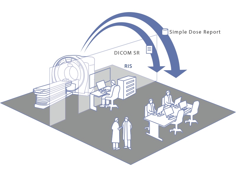
Using the DICOM standard, it is possible to transfer dose information as a DICOM Structured Report (DICOM SR).
Small footprint with 3-unit configuration
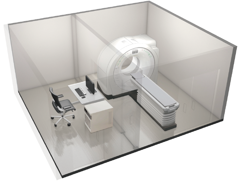
Only the gantry, the patient table, and the operation console configuration*1 is realized. There are no other separate units with build-in system transformer, so the space in the CT room can be used effectively.
- *1 For power supply voltage 200V
Eco mode reduces stand-by power consumption

Supria is equipped with both On-time stand-by and Off-time mode function.
With these Eco mode functions, it reduces power consumption of equipment in the gantry and energization time of the X-ray detector, thereby reducing power consumption during stand-by.
Additional information
| Weight | 1000 kg |
|---|
Images
Intelli IP Imaging
Lowered arms / Pneumonia

Obese patient (Weight : 120kg)


High-Speed Scanning
Chest scanning

Thoracoabdominal scanning

Submillimeter slice imaging for high resolution and high quality images
Scaphoid bone fracture


High quality imaging with the gantry tilt
Cerebral infarction


Lacunar infarction


Clinical images with Intelli IP
Head routine


Pulmonary emphysema


Breast cancer (suspected lymph node metastasis)


Uterine fibroids


Spondylolysis (10 years old )


Cuboid bone fracture


Tibia fracture

Rib fracture


Compression fracture

Abdominal CTA (100kV)


Hepatic hemangioma (Dynamic)


Cerebral aneurysm


Right middle cerebral artery stenosis

SFA total occlusion


Colonic polyp


Images for Veterinary Customers
 Supria In-Room CT Vet Images
FAQ
How does HiMAR work?
HiMAR (High Quality Metal Artifact Reduction) technology in the Fujifilm Supria 32-slice CT scanner works by minimizing artifacts caused by metal objects within the scanned area. These artifacts can distort the image quality and make it difficult to accurately diagnose conditions. HiMAR uses advanced algorithms to differentiate between metal and surrounding tissues, effectively reducing the streaks and distortions that metal objects typically cause
Model-Based Iterative Reconstruction (MBIR) differs from traditional methods like Filtered Back Projection (FBP) and standard Iterative Reconstruction (IR) in several key ways:
- Noise Reduction: MBIR significantly reduces image noise compared to FBP and standard IR. It uses advanced statistical models to iteratively refine the image, resulting in clearer and more accurate images
- Artifact Reduction: MBIR is particularly effective at minimizing artifacts, such as streaks caused by metal objects, which are common in FBP. This leads to more reliable diagnostic images.
- Image Quality: MBIR provides superior image quality with higher spatial resolution and better contrast-to-noise ratio (CNR). This is achieved through its detailed modeling of the imaging system and the scanned object
- Computational Demand: MBIR requires more computational power and time compared to FBP and standard IR. This is due to its complex iterative process, which involves multiple cycles of forward and backward projections to refine the image
- Radiation Dose: MBIR allows for lower radiation doses while maintaining high image quality. This is beneficial for patient safety, especially in repeated imaging scenarios
Overall, MBIR offers significant improvements in image quality and diagnostic accuracy, albeit with higher computational requirements
Image reconstruction in CT scanning is a mathematical process that converts raw data from X-ray projections into detailed cross-sectional images. This process involves several key techniques:
- Filtered Back Projection (FBP): This is one of the most commonly used methods. It involves filtering the projection data to reduce blurring and then back-projecting it to form an image. FBP is known for its computational efficiency and stability
- Iterative Reconstruction (IR): This method improves image quality by iteratively refining the image. It starts with an initial guess and repeatedly adjusts it to minimize the difference between the measured and calculated data. IR can significantly reduce noise and artifacts, leading to clearer images
- Analytical Reconstruction: This category includes methods like FBP and others that use mathematical formulas to directly compute the image from the projection data. These methods are fast but can be limited by noise and artifacts
- Model-Based Iterative Reconstruction (MBIR): This advanced form of IR incorporates detailed models of the imaging system and the scanned object. It provides superior image quality and noise reduction but requires more computational power
These techniques are crucial for producing high-quality images while minimizing radiation exposure to patients
Videos
Recommended products
Nothing found.